Intraductal Oncocytic Papillary Neoplasm of the Pancreas: A Radio-Pathological Case Study
DOI:
https://doi.org/10.6092/1590-8577/3871Keywords:
Carcinoma, Pancreatic Ductal, Diffusion Magnetic Resonance Imaging, Fluorodeoxyglucose F18, Magnetic Resonance Imaging, Pancreas, Positron-Emission Tomography, Tomography, X-Ray ComputedAbstract
Context An intraductal oncocytic papillary neoplasm is a rare pancreatic tumor with the potential of developing invasive carcinoma. Its differentiation from other cystic-like neoplasms of the pancreas, such as intraductal papillary mucinous neoplasms, is a challenge for pancreatic imaging. Case report We present the case of a 76-year-old male with painless jaundice caused by an intraductal oncocytic papillary neoplasm of the pancreas. The imaging findings on computed tomography, magnetic resonance including diffusion-weighted imaging, and 18F-fluorodeoxyglucose positron emission tomography are presented and the radio-pathological correlations are discussed. Conclusion An intraductal oncocytic papillary neoplasm of the pancreas appears as a cystic tumor communicating with the dilated pancreatic duct featuring intraductal tumor nodules. Intraductal oncocytic papillary neoplasms show a high 18F-fluorodeoxyglucose-uptake in positron emission tomography and low diffusion values in diffusion-weighted imaging including apparent diffusion coefficient maps which may be a valuable attribute in distinguishing these rare lesions from intraductal papillary mucinous neoplasms.
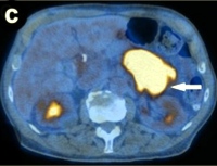
Image: Original attenuation-corrected coronal PET image.
Downloads
References
Adsay NV, Adair CF, Heffess CS, Klimstra DS. Intraductal oncocytic papillary neoplasms of the pancreas. Am J Surg Pathol 1996; 20:980-94. [PMID 8712298] (FULL TEXT: http://journals.lww.com/ajsp/pages/articleviewer.aspx?year=1996&issue=08000&article=00007&type=abstract)
Jyotheeswaran S, Zotalis G, Penmetsa P, Levea CM, Schoeniger LO, Shah AN. A newly recognized entity: intraductal "oncocytic" papillary neoplasm of the pancreas. Am J Gastroenterol 1998; 93:2539-43. [PMID 9860422] (FULL TEXT: http://www.nature.com/ajg/journal/v93/n12/full/ajg1998591a.html)
Nobukawa B, Suda K, Suyama M, Ariyama J, Beppu T, Futagawa S. Intraductal oncocytic papillary carcinoma with invasion arising from the accessory pancreatic duct. Gastrointest Endosc 1999; 50:864-6. [PMID 10570358] (FULL TEXT: http://www.giejournal.org/article/S0016-5107(99)70180-X/fulltext)
Noji T, Kondo S, Hirano S, Ambo Y, Tanaka E, Katoh C, et al. Intraductal oncocytic papillary neoplasm of the pancreas shows strong positivity on FDG-PET. Int J Gastrointest Cancer 2002; 32:43-6. [PMID 12630769] (FULL TEXT: http://www.springerlink.com/content/4002tj83v411h47u/fulltext.pdf)
Oku T, Maeda M, Wada Y, Waga E, Ono K, Nagamachi Y, et al. Intraductal oncocytic papillary neoplasm having clinical characteristics of mucinous cystic neoplasm and a benign histology. JOP. J Pancreas (Online) 2007; 8:206-13. [PMID 17356245] (FULL TEXT: http://www.joplink.net/prev/200703/06.html)
Shima Y, Yagi T, Inagaki M, Sadamori H, Tanaka N, Horimi T, et al. Intraductal oncocytic papillary neoplasm of the pancreas with celiac artery compression syndrome and a jejunal artery aneurysm: report of a case. Surg Today 2005; 35:86-90. [PMID 15622472] (FULL TEXT: http://www.springerlink.com/content/q2w37lku3jwqxq9g/fulltext.pdf)
Thompson K, Castelli MJ, Gattuso P. Metastatic papillary oncocytic carcinoma of the pancreas to the liver diagnosed by fine-needle aspiration. Diagn Cytopathol 1998; 18:291-6. [PMID 9557266] (FULL TEXT: http://www3.interscience.wiley.com/cgi-bin/fulltext/39163/PDFSTART)
Kato Y, Nakagouri T, Konishi M, Takahashi S, Gotoda N, Hasebe T, Kinosita T. Intraductal oncocytic papillary neoplasm of the pancreas with strong accumulation on FDG-PET. Hepatogastroenterology 2008; 55:900-2. [PMID 18705293]
Ishida M, Egawa S, Aoki T, Sakata N, Mikami Y, Motoi F, et al. Characteristic clinicopathological features of the types of intraductal papillary-mucinous neoplasms of the pancreas. Pancreas 2007; 35:348-52. [PMID 18090241] (FULL TEXT: https://journals.lww.com/pages/login.aspx?ReturnUrl=%2fpancreasjournal%2fsecure%2fpages%2fpurchase.aspx%3fan%3d00006676-200711000-00009)
Patel SA, Adams R, Goldstein M, Moskaluk CA. Genetic analysis of invasive carcinoma arising in intraductal oncocytic papillary neoplasm of the pancreas. Am J Surg Pathol 2002; 26:1071-7. [PMID 12170096] (FULL TEXT: http://journals.lww.com/ajsp/pages/articleviewer.aspx?year=2002&issue=08000&article=00014&type=abstract)
Hruban R, Pitman MB, Klimstra DS. AFIP Atlas of Tumor Pathology. Tumors of the Pancreas. Series 4, Fasc 6: Washington, American Registry of Pathology in collaboration with Armed Forces Institute of Pathology, 2007, pp 23-50.
Itai Y, Minami M. Intraductal papillary-mucinous tumor and mucinous cystic neoplasm: CT and MR findings. Int J Gastrointest Cancer 2001; 30:47-63. [PMID 12489580] (FULL TEXT: http://www.springerlink.com/content/f6k23232g4876178/fulltext.pdf)
Strobel K, Heinrich S, Bhure U, Soyka J, Veit-Haibach P, Pestalozzi BC, et al. Contrast-enhanced 18F-FDG PET/CT: 1-stop-shop imaging for assessing the resectability of pancreatic cancer. J Nucl Med 2008; 49:1408-13. [PMID 18703604] (FULL TEXT: http://jnm.snmjournals.org/cgi/content/full/49/9/1408)
Yoshioka M, Sato T, Furuya T, Shibata S, Andoh H, Asanuma Y, et al. Positron emission tomography with 2-deoxy-2-[(18)F] fluoro- d-glucose for diagnosis of intraductal papillary mucinous tumor of the pancreas with parenchymal invasion. J Gastroenterol 2003; 38:1189-93. [PMID 14714260] (FULL TEXT: http://www.springerlink.com/content/5j1lm8h61w81rfuc/fulltext.pdf)
Sperti C, Bissoli S, Pasquali C, Frison L, Liessi G, Chierichetti F, Pedrazzoli S. 18-fluorodeoxyglucose positron emission tomography enhances computed tomography diagnosis of malignant intraductal papillary mucinous neoplasms of the pancreas. Ann Surg 2007; 246:932-7. [PMID 18043094] (FULL TEXT: http://journals.lww.com/annalsofsurgery/pages/articleviewer.aspx?year=2007&issue=12000&article=00004&type=abstract)
Blake MA, McKernan M, Setty B, Fischman AJ, Mueller PR. Renal oncocytoma displaying intense activity on 18F-FDG PET. AJR Am J Roentgenol 2006; 186:269-70. [PMID 16357422] (FULL TEXT: http://www.ajronline.org/cgi/content/full/186/1/269)
Kim DJ, Chung JJ, Ryu YH, Hong SW, Yu JS, Kim JH. Adrenocortical oncocytoma displaying intense activity on 18F-FDG-PET: a case report and a literature review. Ann Nucl Med 2008; 22:821-8. [PMID 19039562] (FULL TEXT: http://www.springerlink.com/content/165g1u00813754x1/fulltext.pdf)
Subramaniam RM, Durnick DK, Peller PJ. F-18 FDG PET/CT imaging of submandibular gland oncocytoma. Clin Nucl Med 2008; 33:472-4. [PMID 18580232] (FULL TEXT: http://journals.lww.com/nuclearmed/pages/articleviewer.aspx?year=2008&issue=07000&article=00005&type=abstract)
Hagino K, Tsunoda A, Ishihara A, Kishimoto S, Suzuki T, Hara A. Oncocytoma in the parotid gland presenting a remarkable increase in fluorodeoxyglucose uptake on positron emission tomography. Otolaryngol Head Neck Surg 2006; 134:708-9. [PMID 16564402] (FULL TEXT: http://www.otojournal.org/article/PIIS0194599805004031/fulltext)
Vilanova JC, Barcelo J. Diffusion-weighted whole-body MR screening. Eur J Radiol 2008; 67:440-7. [PMID 18430538] (FULL TEXT: http://www.ejradiology.com/article/PIIS0720048X08001733/fulltext)
Bruegel M, Rummeny EJ. Hepatic metastases: use of diffusion-weighted echo-planar imaging. Abdom Imaging 2009; May 27. [PMID 19471997] (FULL TEXT: http://www.springerlink.com/content/q0h7353082167771/fulltext.html)
Matsuki M, Inada Y, Nakai G, Tatsugami F, Tanikake M, Narabayashi I, et al. Diffusion-weighted MR imaging of pancreatic carcinoma. Abdom Imaging 2007; 32:481-3. [PMID 17431713] (FULL TEXT: http://www.springerlink.com/content/08714340n8032234/fulltext.html)
Takeuchi M, Matsuzaki K, Kubo H, Nishitani H. High-b-value diffusion-weighted magnetic resonance imaging of pancreatic cancer and mass-forming chronic pancreatitis: preliminary results. Acta Radiol 2008; 49:383-6. [PMID 18415779] (FULL TEXT: http://informahealthcare.com/doi/full/10.1080/02841850801895381)
Yamashita Y, Namimoto T, Mitsuzaki K, Urata J, Tsuchigame T, Takahashi M, Ogawa M. Mucin-producing tumor of the pancreas: diagnostic value of diffusion-weighted echo-planar MR imaging. Radiology 1998; 208:605-9. [PMID 9722835] (FULL TEXT: http://radiology.rsna.org/content/208/3/605.long)
Kartalis N, Lindholm TL, Aspelin P, Permert J, Albiin N. Diffusion-weighted magnetic resonance imaging of pancreas tumours. Eur Radiol 2009; 19:1981-90. [PMID 19308414] (FULL TEXT: http://www.springerlink.com/content/m3310p70g6121thw/fulltext.html)
Ozsunar Y, Sorensen AG. Diffusion- and perfusion-weighted magnetic resonance imaging in human acute ischemic stroke: technical considerations. Top Magn Reson Imaging 2000; 11:259-272. [PMID 11142625] (FULL TEXT: https://journals.lww.com/topicsinmri/secure/pages/purchase.aspx?an=00002142-200010000-00003)
Inan N, Arslan A, Akansel G, Anik Y, Demirci A. Diffusion-weighted imaging in the differential diagnosis of cystic lesions of the pancreas. AJR Am J Roentgenol 2008; 191:1115-21. [PMID 18806153] (FULL TEXT: http://www.ajronline.org/cgi/content/full/191/4/1115)
Naganawa S, Kawai H, Fukatsu H, Sakurai Y, Aoki I, Miura S, et al. Diffusion-weighted imaging of the liver: technical challenges and prospects for the future. Magn Reson Med Sci 2005; 4:175-186. [PMID 16543702] (FULL TEXT: http://www.jstage.jst.go.jp/article/mrms/4/4/175/_pdf)